Cardiovascular sonography is the highest paying sonography specialty today, earning up to $100,000 annually in certain parts of the United States. Remember that sonographers who specialize in the cardiovascular system are different from cardiac technicians; CV sonographers are diagnostic medical sonographers who are specialized in performing sonography on the heart and the rest of the circulatory system.
What does a sonographer do?
|
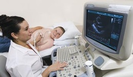
Sonographers are diagnostic technicians who are trained to use machines called sonograms. Sonograms send high-frequency sound waves through the body, which bounces off of solid structures like bones and organs. Sonographers, like all other allied health professionals, are to become specialized, study specialty sonography for different parts or systems of the body.
How to become a specialty ultrasound technician?
|
|
As of 2014, the Commission on Accreditation of Allied Health Education Programs (CAAHEP) recognizes four specialties that can have their own programs: general, cardiac, vascular, and pediatric sonography. There are 211 schools in the US who offer ultrasound technician programs that are accredited by the CAAHEP. If you are a sonographer who wants to specialize in cardiovascular sonography, the top schools that you should enroll in are CAAHEP-accredited colleges.
Where is the best place to work at as a sonographer?
Currently, the top paying state and the state with the highest employment levels for sonographers is California. In California, there are 4,710 sonographers employed who earn $41.61hourly, amounting to an annual salary of $86,550. There are certain areas in California that offer even higher pay, particularly the San Francisco-San Mateo-Redwood metropolitan area where sonographers earn $111,700 annually – the highest paying area in the country. The CAAHEP currently has 10 accredited ultrasound technician schools in California, seen through the link.
Learning specialty skills in cardiovascular sonography
One of the skills that a sonographer learns when he or she specializes in cardiovascular sonography is guided central venous catheter insertion. Approximately 25000 CVCs are inserted each year in the UK, and with this incredibly high number, having a clear guide during insertion has been shown to decrease placement failures and damage to the vessel walls. Patient outcomes have been observed to be improved if sonography is used to guide the placement as well as assess it once the catheter is in place.
A study in 2006 revealed that there was a decrease in the frequency of carotid artery puncture and hematoma when sonography was used during the insertion. Further studies have actually shown an increase in first-puncture and successful catheter insertion with sonography. These are very compelling in the benefits of CVC-insertion skills in cardiovascular sonography.
A study in 2006 revealed that there was a decrease in the frequency of carotid artery puncture and hematoma when sonography was used during the insertion. Further studies have actually shown an increase in first-puncture and successful catheter insertion with sonography. These are very compelling in the benefits of CVC-insertion skills in cardiovascular sonography.
Proper training in sonography
Because sonography is a relatively new technique to other allied health professionals who are allowed to insert CVCs, intensive training is required. While sonographers can be trained to perform the insertion themselves, other professionals on the other hand can be trained to use sonograms as well. Another study done by Bold, et. al. in 1998 recommend that professionals practice with training models to assure themselves as well as the patients of the CVC-insertion skill they just learned.
