The WHO and SonographY
|
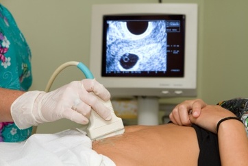
Because sonography is a diagnostic medical procedure, the World Health Organization has come up with guidelines on how to perform it correctly. The general consensus is that sonography is one of the safest procedures used on patients today. It poses no threat to people who are immunocompromised or pregnant – making it quite safe for expecting mothers and patients with cancer or similar diseases.
Sonographers have become one of the most popular medical careers to date, with employment levels expected toincrease by 46 percent between 2012 and 2020. You can find complete (Infographic: Employment and Salary Statistics for Sonographers through the link. The WHO has created a manual of diagnostic ultrasound in order to help guide health care providers performing the procedure. |
|
Basic physics of ultrasound
When sound is used at frequencies of 20,000 Hertz (vibrations that cannot be heard by the human ear), it is called an ultrasound – the term where sonography gets its name from. Sonography is typically given at frequencies of 1 to 30 Megahertz. The procedure is itself is depends on the analysis of these sound waves, done by a computer or a similar machine. It is only after the images have been created that the sonographer can analyze the results and make a report.
Generating an ultrasound
Because ultrasounds are very high frequencies, it takes special equipment and materials to generate them. Piezoelectric crystals are used to convert mechanical pressure in an electric voltage on their surface. The alternating electric voltage causes oscillations or vibrations (the “ultrasound”) which can be sent out by a transducer that is placed on the body. Transducers are thin discs made of ceramic material, whose thickness determines the ultrasound’s frequency. These discs are very thin, usually less than 1 mm thick.
The acoustic shadow
One of the major characteristics of a diagnostic medical ultrasound is the acoustic shadow. The images produced by sound waves are black and white images, casting off “shadows” that are detected by the high frequencies. When sound bounces off a solid structure, it creates an image that is seen on the monitor, differentiated from the surrounding space by a shadow. The higher the frequency, the better the quality and resolution of the image.
Doppler effect
First postulated by Christian Doppler, the Doppler Effect is used to create images of moving objects – used specifically for creating images of blood flow through vessels and tissues in the body. The effect is made possible because frequencies are higher as a moving object approaches and lower as the moving object moves away. This difference in frequency is called the Doppler frequency, and is responsible for the images that are captured using a Doppler machine (a variation of a sonogram).
Ultrasound techniques
Like a Doppler ultrasound, there are different ultrasound techniques that are employed in medical diagnostics. The A-mode is a one-dimensional technique that used one crystal. Because one-dimensional techniques are very limited in terms of what information can be taken from the results, it is rarely used today.
B-mode (brightness modulation) is the more popular choice, because it able to produce grayscale images according to the signal’s intensity. A variation of this is the B-flow technique (three dimensional), able to visualize blood flow without using the Doppler technique. M-mode or TM-mode (time motion) is also used to analyze moving structures, such as the valves of the heart. TM-mode ultrasounds are simply B-ultrasounds that are recorded continuously over time. The year 2016 sees more advancement made to the field of sonography, spearheaded by the WHO.
