|
Echocardiograms are one of the most popular sonogram procedures performed in hospitals and diagnostic laboratories. Like other sonogram procedures, an echocardiogram uses sound waves to create medical images of the heart and its structures. Echocardiograms come in 2-dimensional, 3-dimensional, and Doppler ultrasound forms, used in routine diagnostic procedures for cardiovascular disease and otherwise.
Sonographer specialties
When a person trains to be a sonographer, he or she chooses a specialty immediately after starting training or on-the-job. There are four major specialties recognized by the Commission on Accreditation of Allied Health Education Programs (CAAHEP) that can have their own Diagnostic Medical Sonography (DMS) program: general, cardiac, vascular, and pediatric cardiac. Other specialties are included in general training, like abdominal and obstetric and gynecologic sonography.
|
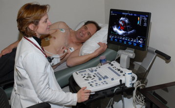
Transthoracic Echocardiograms (TTE)
|
How much do ultrasound techs make in 2014 is a common factor considered by most students who are considering to become an ultrasound tech. Salary averages around $67,000 per year in the US for radiology technicians, high compared to other medical professionals. The predicted increase in employment and salary is actually 46 percent between 2012 and 2022 – higher than the nationwide average 11 percent for all occupations.
Transthoracic (TT) and transesophageal (TE) echocardiograms
Echocardiograms can be given in two ways: one non-invasive and one invasive method. The non-invasive method is called transthoracic (TT) echocardiogram, the more popular method used in routine examinations. Because TE echocardiograms are invasive, they are only used to confirm diagnoses because they provide much clearer images of the heart. When giving a sonogram, a transducer is placed on the area that needs to be examined and sends out sound waves that pass through the body.
In a TT echocardiogram, the transducer is placed on the left side of the chest, directed towards the heart. The transducer is positioned on the left side of the breast bone, left to the nipple, and from the upper abdomen. The sound waves are sent through the body and return to the sonogram as electrical impulses. The impulses are converted into moving and still images of the heart.
The same process is used in a TE echocardiogram, but the transducer is inserted into the esophagus using a scope until it reaches a certain depth to create clear images of the heart.
Use of contrast
The use of contrast is a common practice in sonography. Contrast is injected intravenously (IV), and is used in order to clearly differentiate tissue from blood. Blood appears black on 2D-echocardiography results because the sound waves are scattered by red blood cells, too weak to be picked up by the sonogram. Contrast echocardiography is able to improve the ability of the sonogram to recognize blood pooling and blood/tissue interface – improving the quality of the images of the heart and blood flow through the heart.
Training on-the-job
Sonography is a medical profession where much of the skills are learned on-the-job. When you study in an accredited educational institution, you will most likely have clinical training whether you study on-campus or on-line. Most health care institutions also offer on-the-job training which count as CME units (continuing medical education) for sonography. Choosing the best under/postgraduate course is the first part in a successful career as a sonographer.
