B-Mode Ultrasound Innovations
Innovations made to ultrasound have drastically improved over the years. Learn about B-mode ultrasound scanning and its applications today.
Since Dr. Karl Dussik’s application of ultrasound in medical diagnostics, sonography today is an institution all on its own. It is a widely used procedure for a variety of cases because it is cost-effective as it is an accurate diagnostic measure. Today, ultrasounds are portable – some transducers function simply attached to a tablet or a netbook (a small laptop) – but doesn’t compromise the quality of images that can be taken by the machine.
A kind of ultrasound – the B-Mode Ultrasound – has seen numerous innovations made to the procedure. For a sonographer who is constantly seeking to improve his or her sonography practice and career, training to learn these new skills and techniques in ultrasound technology should be a priority. A high salary comes with the territory when you work in a medical career, but pursuing higher education should always be first on the list (for more details regarding ultrasound tech salary, visit the link).
A kind of ultrasound – the B-Mode Ultrasound – has seen numerous innovations made to the procedure. For a sonographer who is constantly seeking to improve his or her sonography practice and career, training to learn these new skills and techniques in ultrasound technology should be a priority. A high salary comes with the territory when you work in a medical career, but pursuing higher education should always be first on the list (for more details regarding ultrasound tech salary, visit the link).
|
The basic principles of sonography
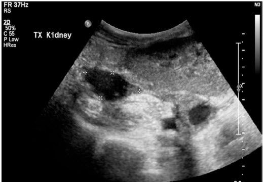
The principles used in sonography are quite simple. Sound waves are created in the transducer and sent through the body. They bounce off of solid structures and tissues and return to the machine, creating images of the body’s internal structures. B-mode stands from brightness-mode, which uses a pulse-echo approach. Sound waves are sent in small pulses, creating a 2-D image. The sound waves are created through the use of piezoelectric crystals, which are thin sheets that vibrate and create sound when an electric current is applied. This is called the piezoelectric effect. Depending on the amount of electricity used, the sheets can vibrate at a higher or lower frequency. Some of the sound waves bounce back while some are absorbed by tissue and transformed into heart. |
Innovations in B-mode ultrasound
Tissue harmonic imaging and spatial compound imaging are two new innovations made to B-mode sonography. Tissue harmonic imaging refers to sound waves that are sent in a single transmission whose frequencies increase or decrease by integral multiples of the original transmission. As the waves pass through tissue, they become distorted or “sharper”. This technique is very popular because it is able to create images beneath deep tissue, or patients with thick and complicated wall structures.
Spatial compound imaging is also known as multibeam imaging. It uses sound waves that are steered (electrically) to create an image of a single area or tissue using different beams. This produces multiple images of a single area, which are then compounded into a single image. The image quality is much better using this technique, with the images appearing less “grainy”.'
Studying CMEs
Currently, there are more than 200 accredited schools in the US that have sonography (Diagnostic Medical Sonography/DMS) programs available to people who want to become an ultrasound tech. Sonographers who are currently working can learn these new skills and techniques through a continuing medical education (CME) program, which can come in the form of seminars, short courses, or research. CMEs are the best ways to improve sonography practice and earn more credentials. B-mode isn’t the only form of sonography that has new innovations and improvements.
Different aspects of the procedure have been generally improved over the years, with sonography being used therapeutically for a variety of conditions. You can inquire about CMEs on technological advancement and career opportunities from different organizations, such as the SDMS or your local college or university.
